
projects


Projects
Polarisation-Sensitive Optical Coherence Tomography
Primary Investigator - Prof. Stephen Matcher
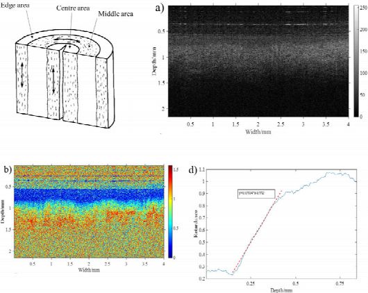
PS-OCT images of human cervical tissue in middle area.
Top-left shows a schematic diagram of the cervix, with the predominant collagen fibre orientation shown by double-headed arrows.
Top-right shows a conventional OCT image, taken from the "middle" area, where pixel brightness represents local optical backscatter intensity, similar to an ultrasound scan. The cervical epithelium is just discernable, as a superficial darker band between depths 0.6 to 0.75 mm. The underlying collagen-rich stroma appears as a brighter area, below this.
Bottom-left shows the same tissue site imaged by PS-OCT. The image now represents optical "retardance", with blue denoting very low retardance and red denoting high retardance. The superficial blue band corresponds to the cervical epithelium, which is composed of tightly packed cells but with little collagen. The increase of retardance with depth into the stroma indicates the presence of aligned collagen fibres.
Bottom-right shows the retardance value plotted as a function of depth. The slope of this line is a potential biomarker of cervical collagen content, which may be predictive of spontaneous pre-term birth. Reproduced from Li et al, "Polarization-Sensitive Optical Coherence Tomography with a conical beam scan for investigation of birefringence and collagen alignment of human cervix", Biomed. Opt. Express 10(8), 4190-4206 (2019).
- Address: The University of Sheffield, Western Bank, Sheffield, South Yorkshire, S10 2TN, UK
- Phone: +44 (0) 114 215 9674
- Email: info@ecclippx.co.uk